Facilities &
Equipment
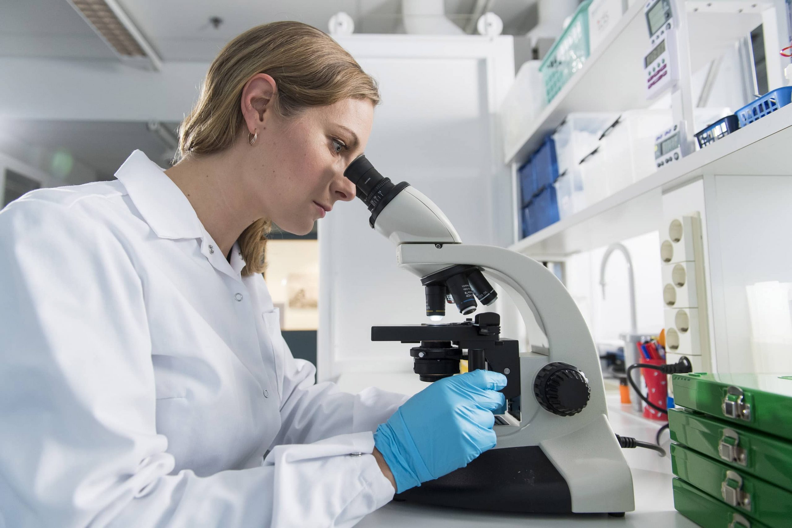
Facilities & equipment
Our preclinical testing facilities are located in Turku, Finland. Our modern facilities include main office, laboratories and animal facilities.
State-of-the-art equipment
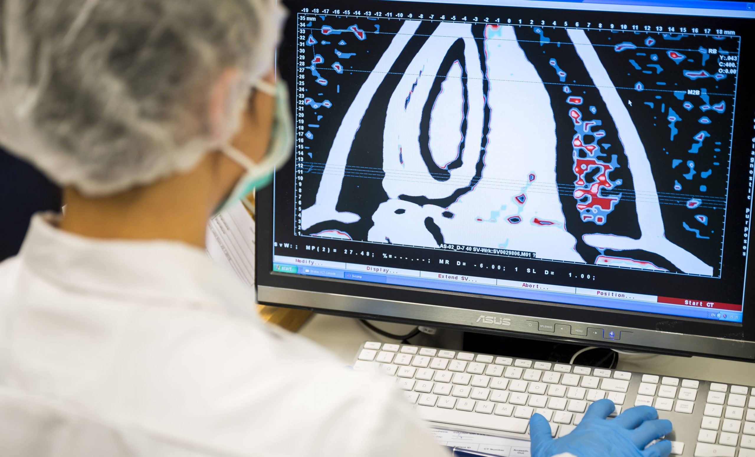
Advanced preclinical imaging solutions are an essential tool for a broad spectrum of application fields such as oncology and skeletal disease models. Our fully equipped laboratory offers on a wide range of specialist equipment to image and study cells and animals on the organ, tissue, cell, or molecular level enabling us to study and monitor the disease progression throughout the study.
High resolution imaging capabilities and speciality in vivo/ex vivo equipment include:
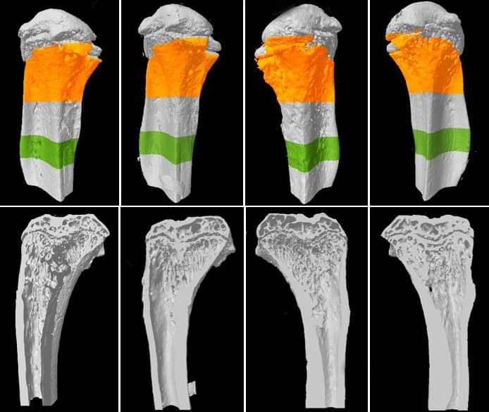
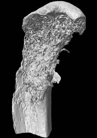
- SkyScan 1276 by Bruker – Ultra-high resolution MicroCT enables 2D/3D image analysis, bone morphology and realistic visualization, full body mouse and rat scanning.
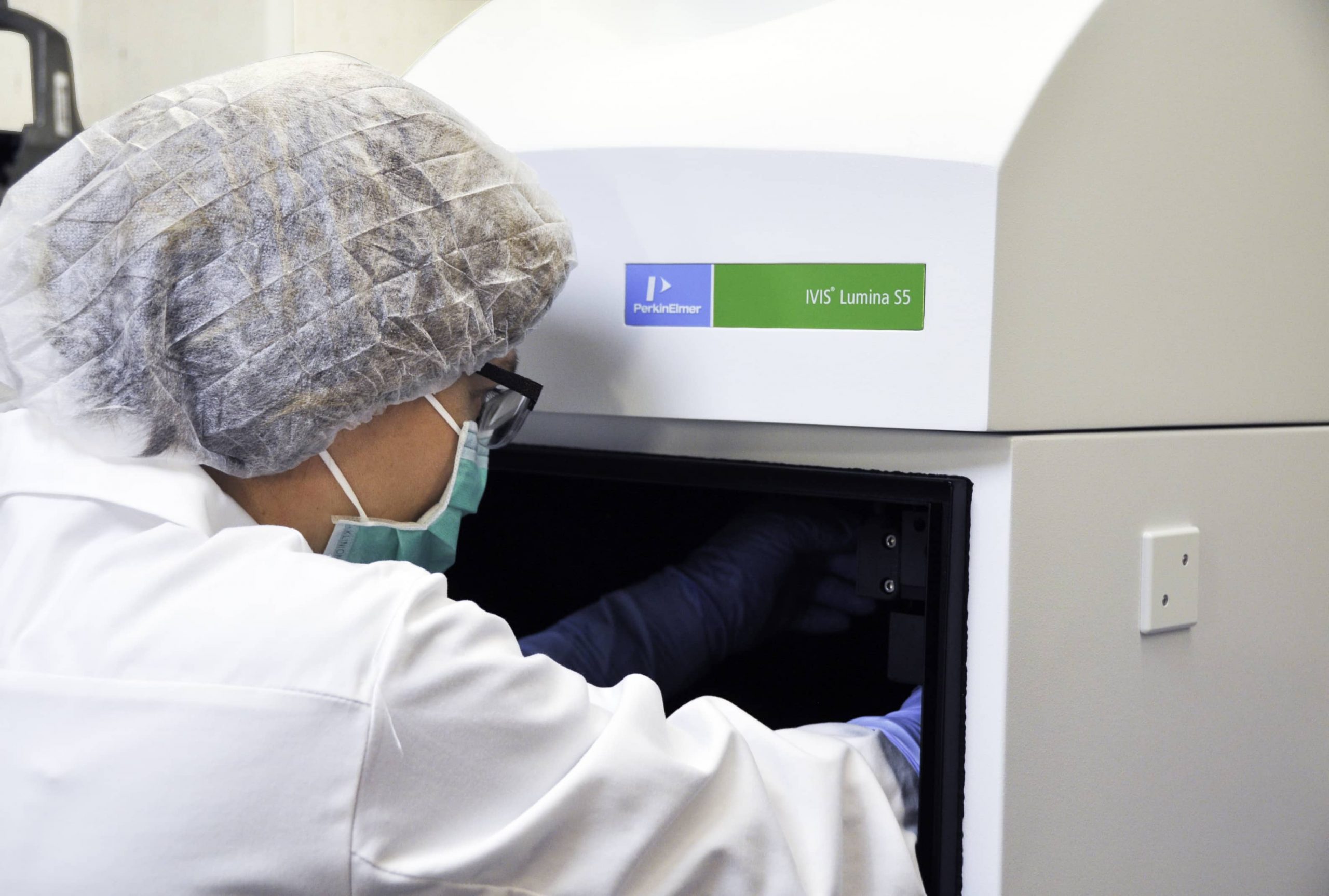
- IVIS Lumina S5 – high-throughput 2D optical imaging system with high-sensitivity bioluminescence and fluorescence
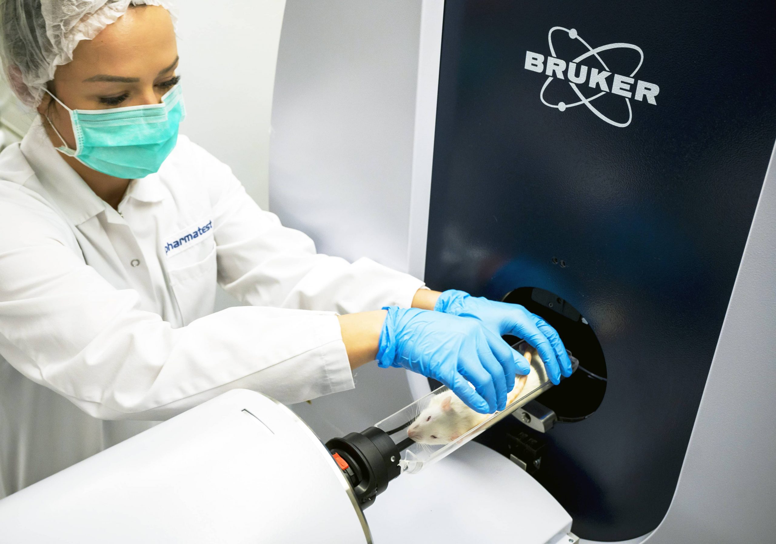
- XCT Research SA+ pQCT – Peripheral quantitative computed tomography (pQCT) for the measurement of small laboratory animals like mice or rats. Evaluation of volumetric bone density, bone geometry and muscle parameter at small laboratory animals like mice and rats using pQCT.
- Faxitron Ultrafocus DXA – fully shielded X-ray cabinet system which allows high resolution 2D X-ray images for small animal imaging.
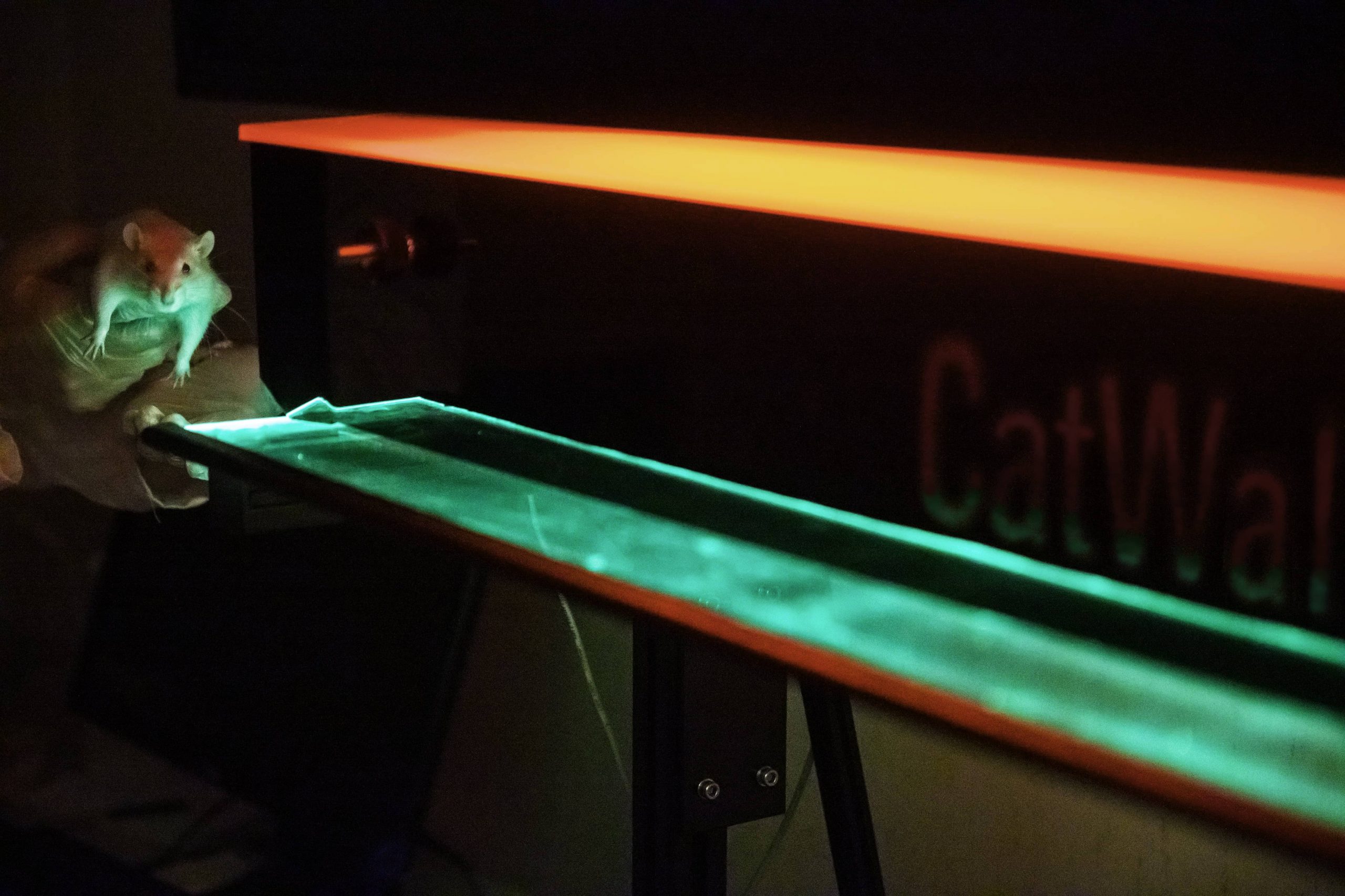
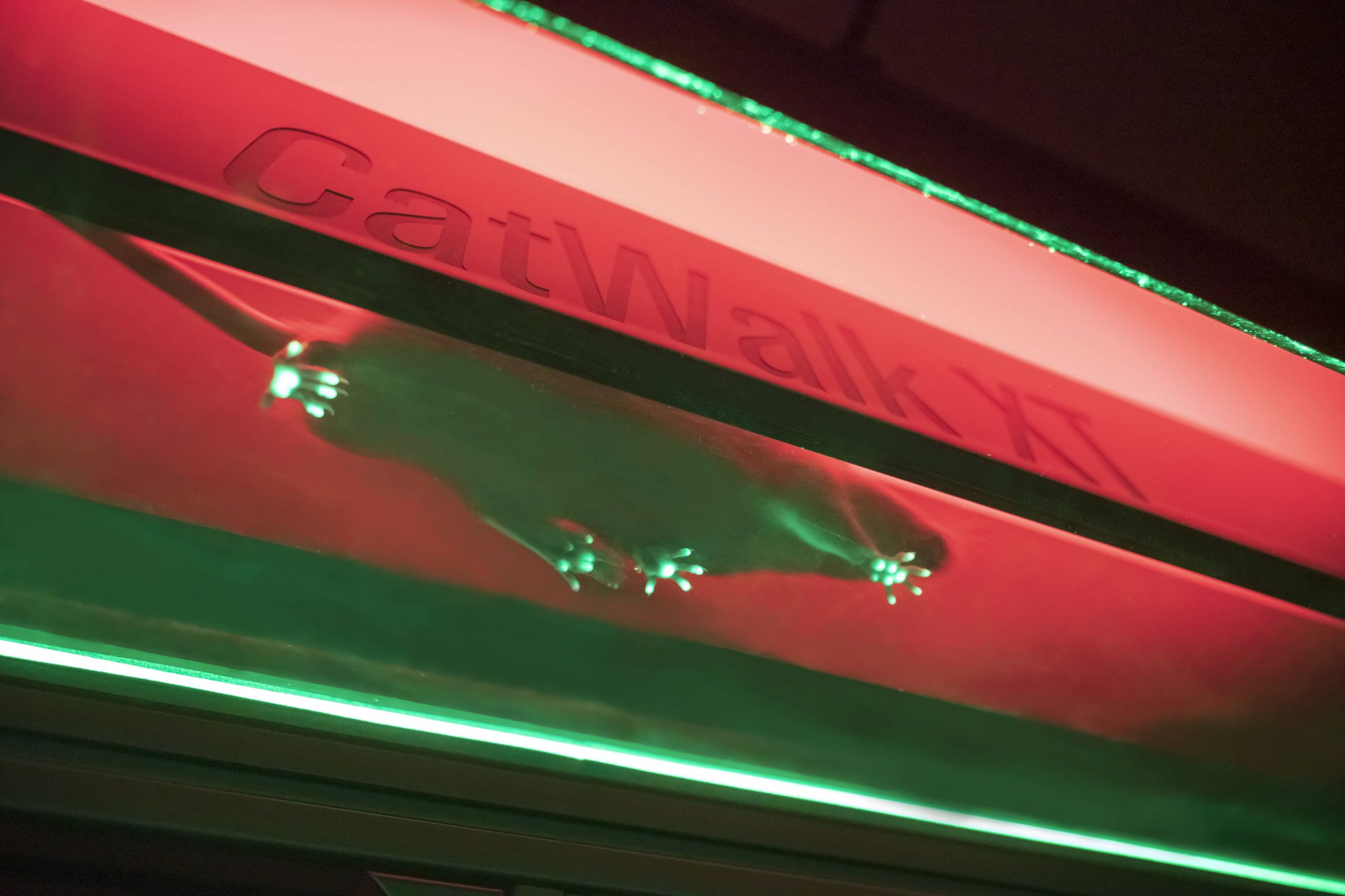
- CatWalk CT by Noldus – detailed analysis system of gait for quantitative assessment of footfalls and locomotion in rats and mice.
Speciality in vitro equipment:
- IDS-iSYS Multi-Discipline Automated System – fully-automated analyzer based on chemiluminescence and absorbance technology for high volume immunoassay testing.
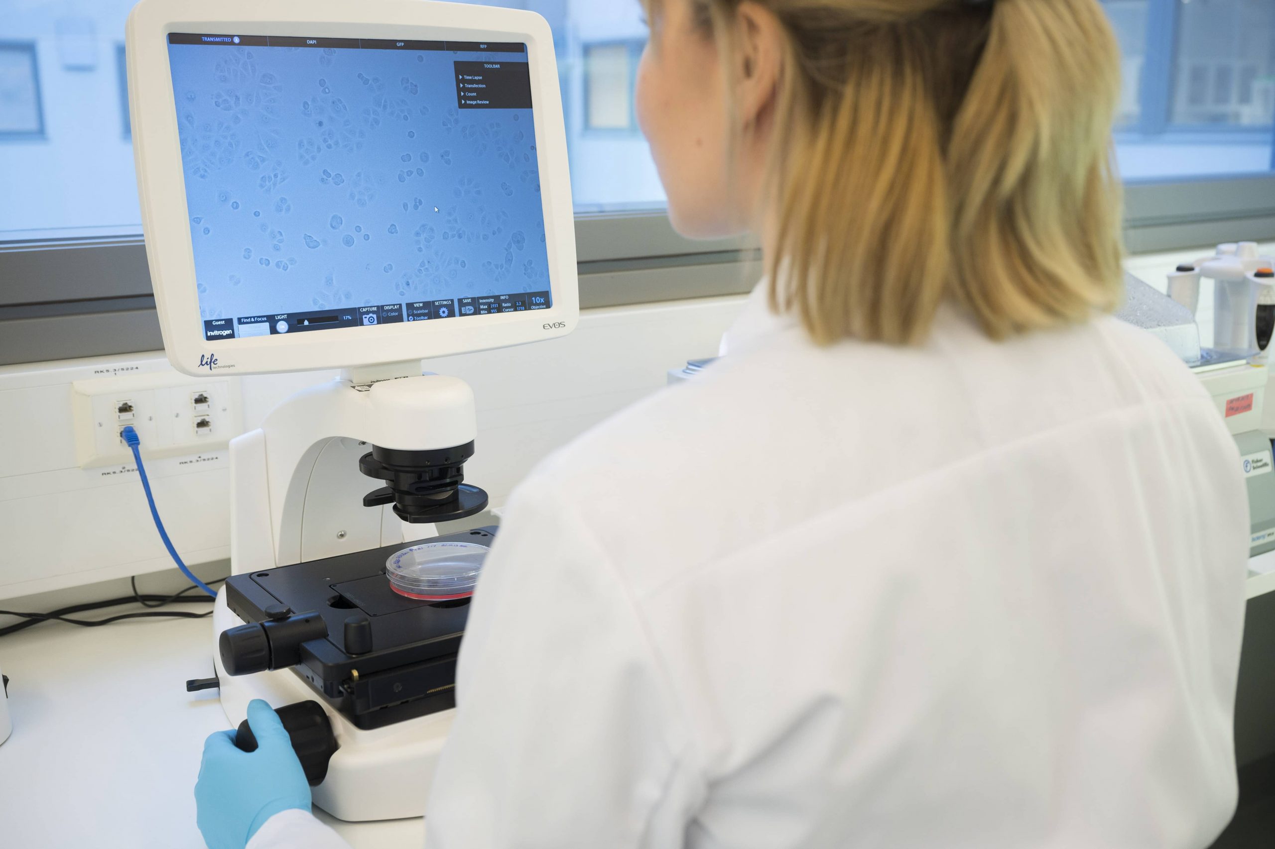
- EVOS cell imaging system – microscope for fluorescence and bright field imaging of cells.
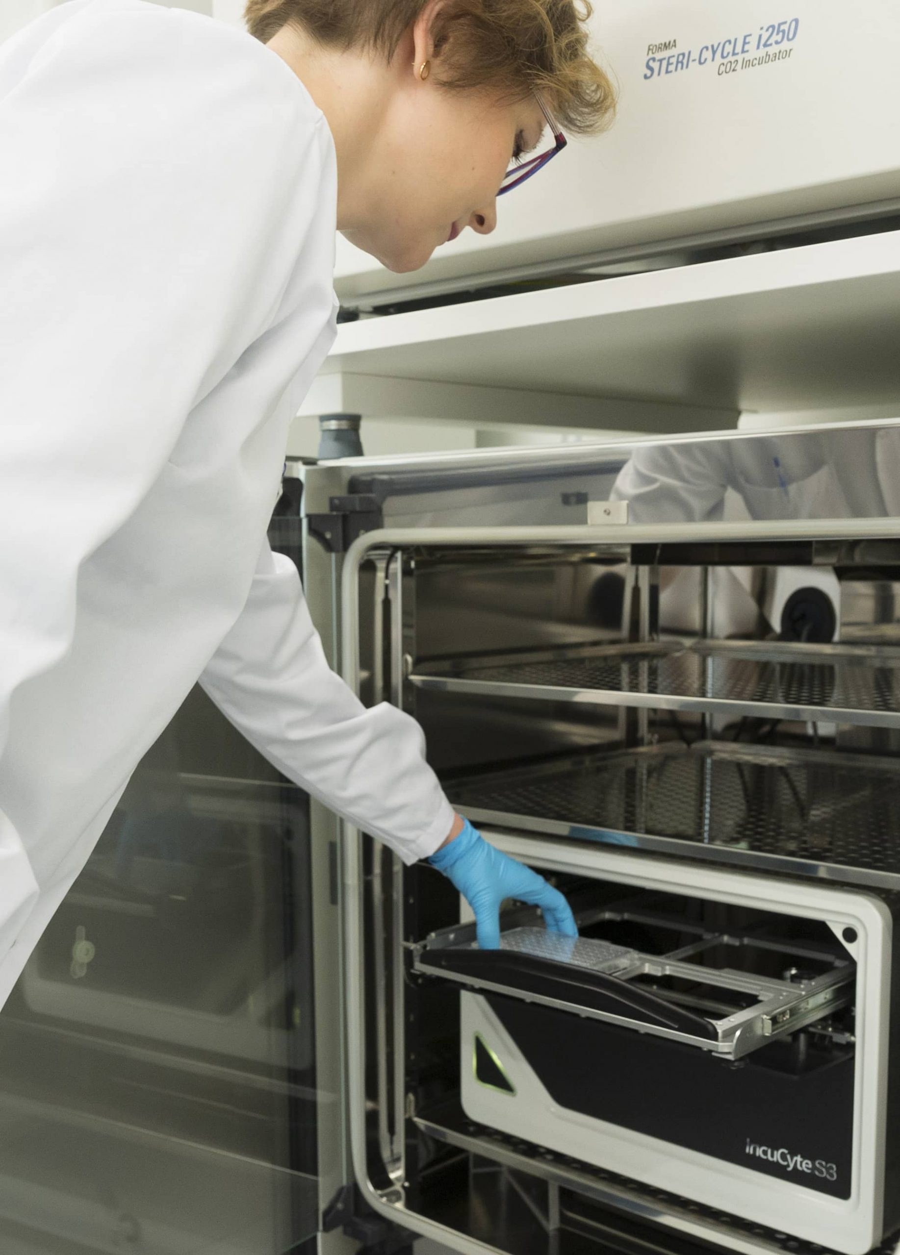
- Incucyte Live-Cell Imager – real-time quantitative image-based analysis with the convenience and throughput of microplate assays that enables visualization and quantification of cell behavior over time.
In vivo laboratories
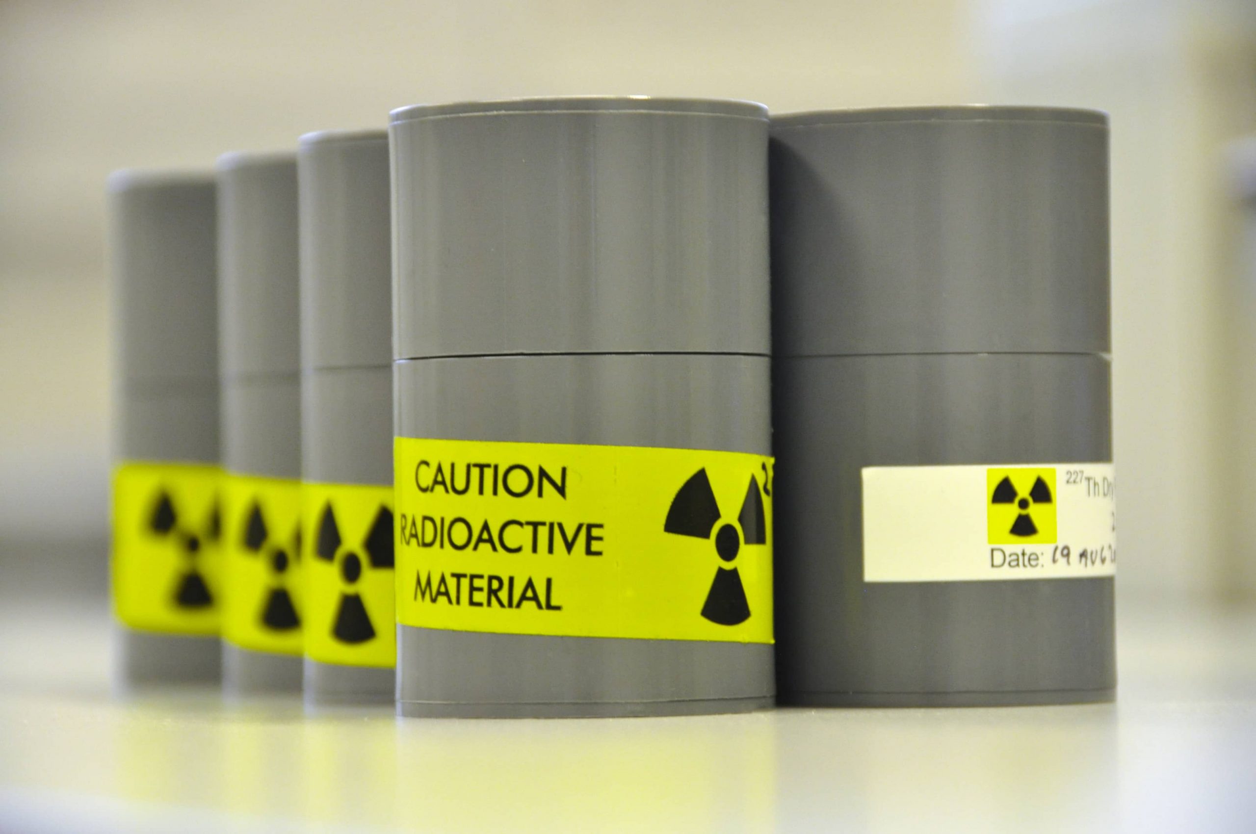
Our animal facility operates in compliance with OECD Principles of Goof Laboratory Practice (GLP). Facilities are inspected regularly by Finnish Medicines Agency and Finnish experimental animal agency’s county veterinarian.
Our animal facilities are inspected and approved by local animal welfare and health authorities. Animal experiments are conducted with the approval of the National Committee for Animal Experiments. We also follow strict guidelines and regulations by Commission on the welfare and use of animals in studies.
We are committed to responsible animal care and provide the highest standards of care and ethical conduct in providing for animal welfare. Environmental conditions of the animal facilities are continuously monitored and controlled.
All animals used in experiments are from commercial breeders. Animals are health checked when they enter our facilities to ensure microbiological quality of laboratory animals. Quarantine and acclimatization periods are mandatory before entering into in-life phase of the experiment. Mice are kept in top-of-the-line individually ventilated cages ensuring high level of bio-containment.
Our animal caregivers have gone through hands-on animal welfare training and have extensive experience with different preclinical models. All animals in our care are provided enriched environment including nesting materials, cage balconies, and cardboard houses.
Principles of the 3R’s
We follow the principle of the 3Rs in our studies: replacement, reduction and refinement. Carefully drafted study designs, statistics and pilot studies to make sure the proper care and use of animals. Continuous monitoring and assessment are done in each study to ensure the wellbeing of animals at all times (in accordance with FELASA requirements.)
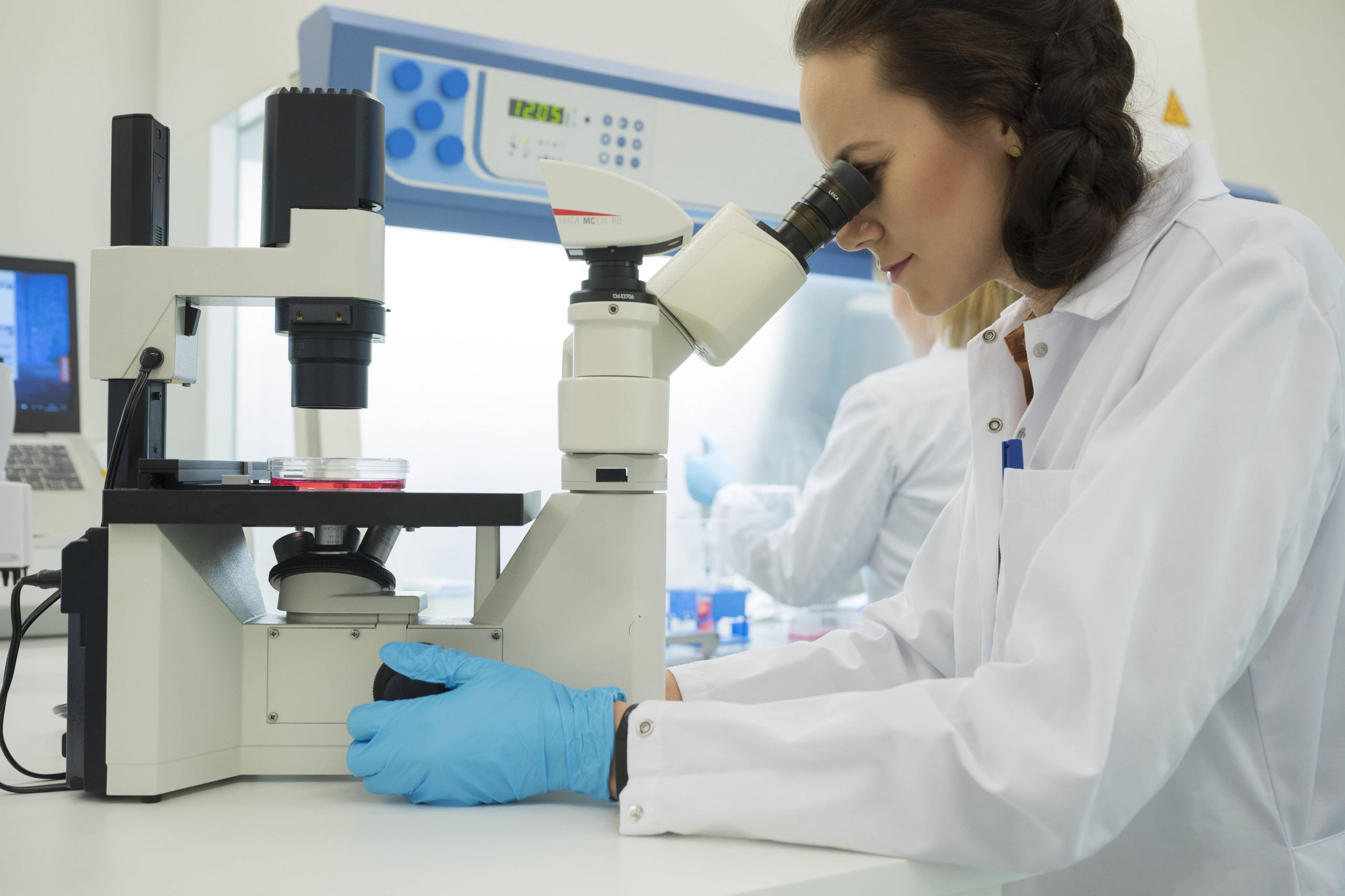
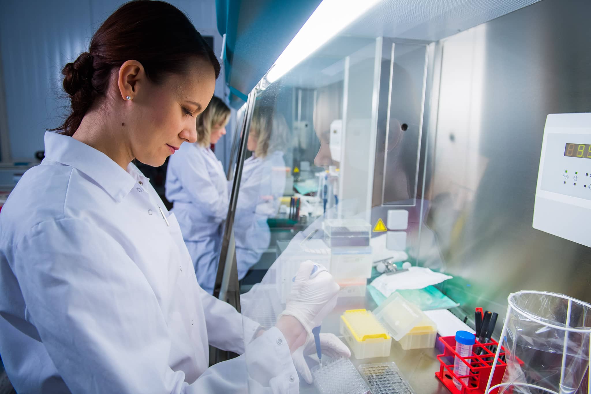
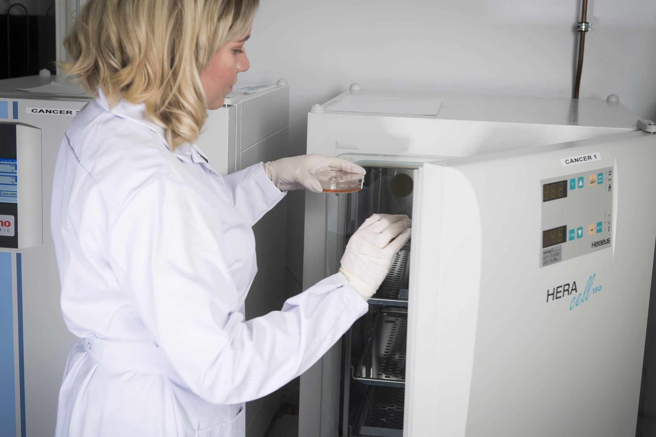
Cell culture
Our facilities include two state-of-the-art cell culture laboratories equipped with multiple incubators and laminar hoods for sterile work. One laboratory is dedicated to primary cell culture and another laboratory is for assays conducted with cell lines. The staff is trained for sterile cell culture work and only the trained personnel has access to the culture spaces.
Our in-house cell line portfolio includes several authenticated cell lines, but we are also cultivating a large variety of cells from our customers. Therefore, we have a very vast experience with different cell types and culture methods. We culture cells also for our in-house in vivo animal experiments and prepare the cells ready to be introduced to the animals. All the cells for animal experiments are tested for pathogens prior to start of the in-life phase.
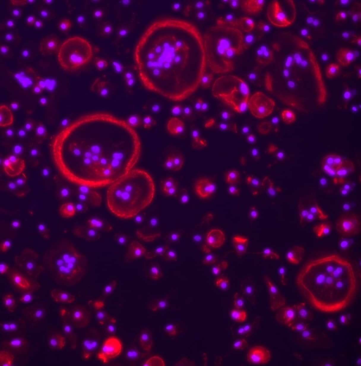
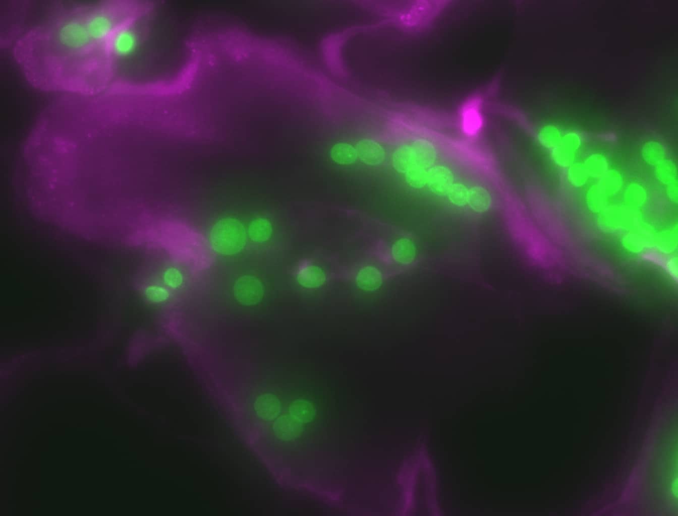
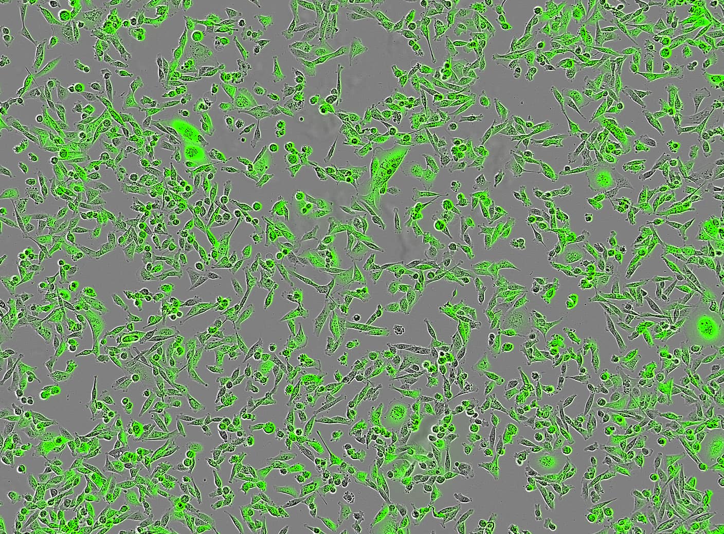
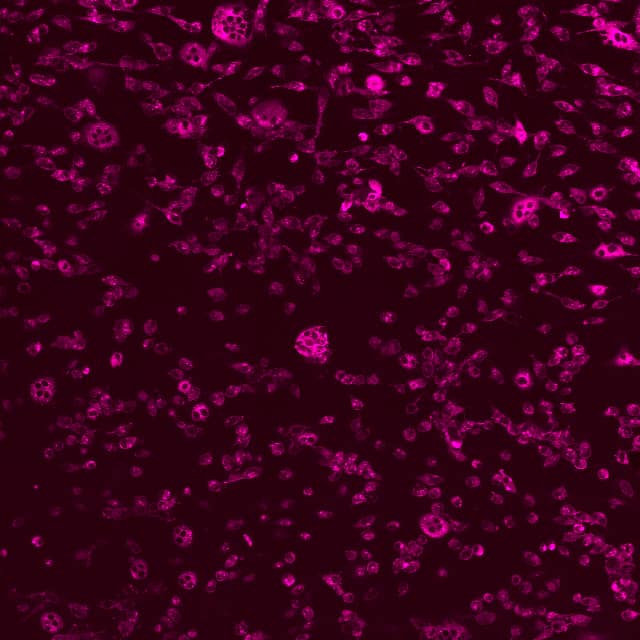
Our cell culture work includes versatile cancer and bone cell assays, from cancer cell migration and viability assays to osteoblast and osteoclast experiments. We also perform co-cultures, for example combining immune cells with cancer cells. While in culture, labelled cells can be monitored with EVOS cell imaging system, and the growth of cells can be observed in real-time with a live cell imager Incucyte. Cells can also be subjected to different stainings after fixation followed by microscopic analysis. Also, expression studies can be performed after termination of cultures.
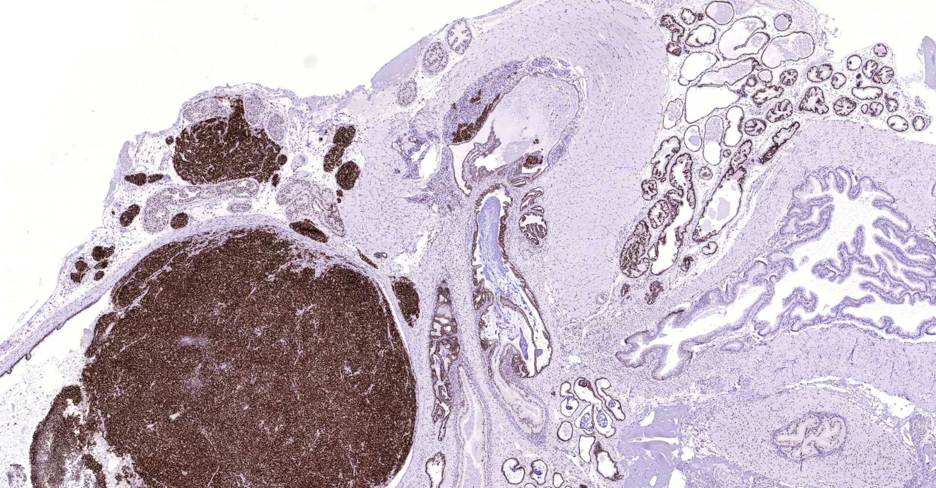
In vitro laboratories
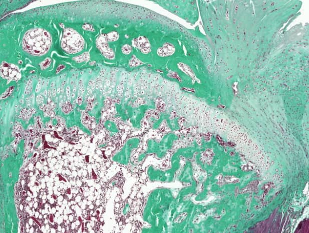
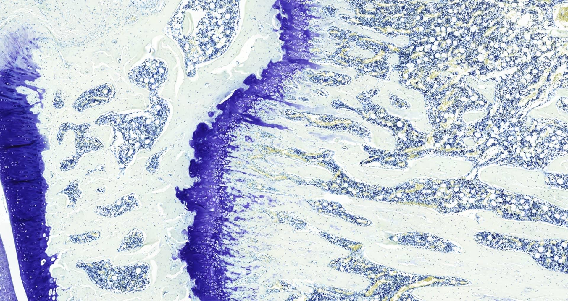
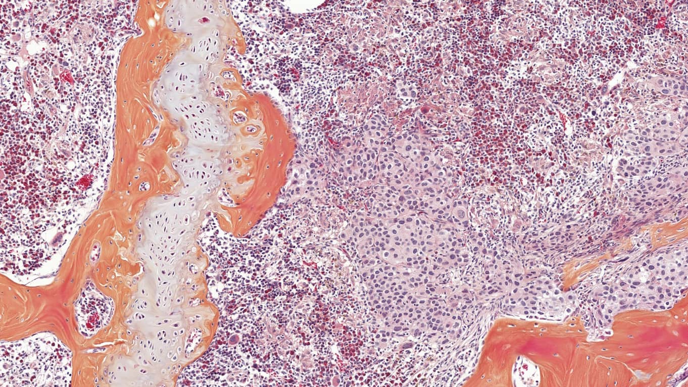
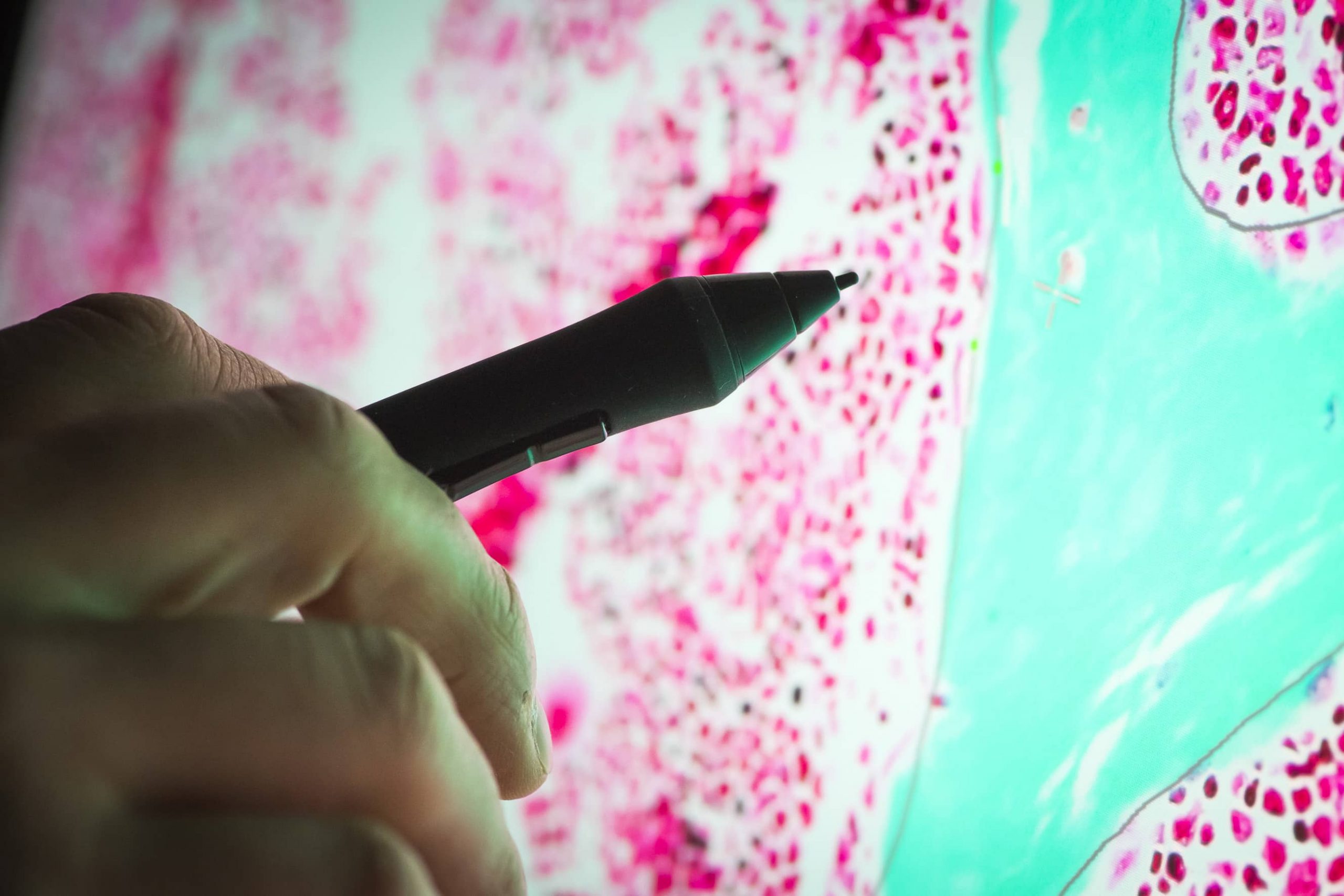
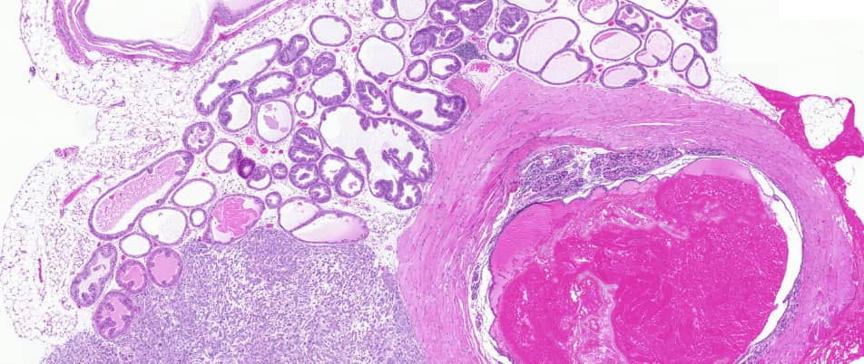
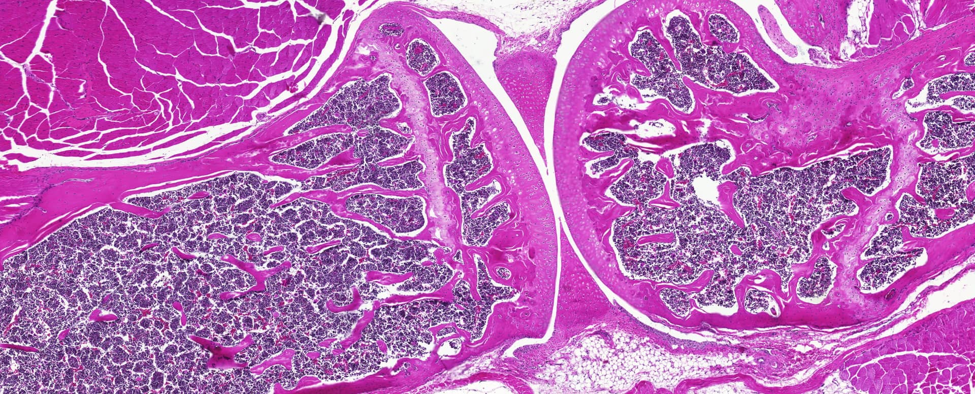
Our in vitro laboratories host several histological and molecular biology activities. We perform immunohistochemistry staining followed by analysis. The analysed tissues come from our in vivo studies or directly from our customers. We have special expertise in bone analysis.
We also perform ELISA analysis of serum, plasma and cell culture samples. Multiplate readers allow measuring several samples in parallel. For example, bone turnover markers can be measured. In addition to samples obtained from animals and cells, we measure markers of clinical blood samples with IDS iSYS Multi-Discipline Automated System that allows automatic record of each sample and thus helps to accurately track even big number of samples.
Samples for different expression analysis are also prepared in-house. We can stain cell or tissue samples for flow cytometric analysis and extract RNA for further expression studies.